DXA is a cost-effective and widely available imaging method used worldwide to better understand osteoporosis and other diseases and treatments that affect bone health. Due to its low cost and minimal radiation dose, DXA is ideally positioned to enable researchers to recruit large numbers of patients and perform multiple follow-up examinations in a short time frame.
Despite its many advantages, DXA also has limitations, as it only provides a two-dimensional assessment of bone mineral density, known as areal bone mineral density (aBMD). This two-dimensional representation of a three-dimensional structure, while good, limits the possibility of a deeper understanding of bone disease, as key data on the condition and response of the cortical and trabecular bone compartments are missing.
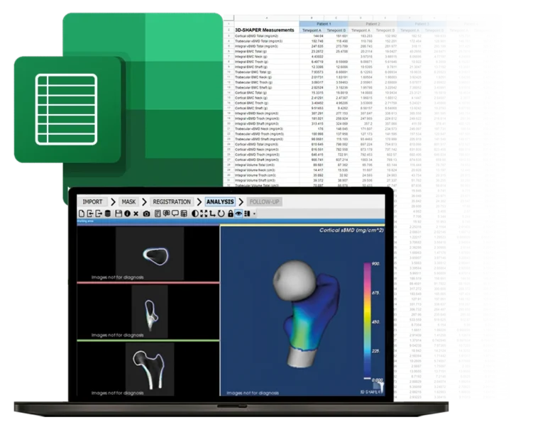
What if you could obtain quantitative computed tomography (QCT)-like data from a routine DXA scan? Well, now it’s possible with 3D-Shaper® Research software, an innovative solution that bridges the gap between DXA-based and QCT assessments by providing patient-specific, QCT-like three-dimensional analysis from a standard two-dimensional DXA image. This means more patients, more time points, and easier Institutional Review Board (IRB) approvals – all for a fraction of the cost.
This website uses cookies to enhance your experience. Some are essential for site functionality, while others help us analyze and improve your usage experience. Please review your options and make your choice.
If you are under 16 years old, please ensure that you have received consent from your parent or guardian for any non-essential cookies.
Your privacy is important to us. You can adjust your cookie settings at any time. For more information about how we use data, please read our privacy policy. You may change your preferences at any time by clicking on the settings button below.
Note that if you choose to disable some types of cookies, it may impact your experience of the site and the services we are able to offer.
Some required resources have been blocked, which can affect third-party services and may cause the site to not function properly.
This website uses cookies to enhance your browsing experience and ensure the site functions properly. By continuing to use this site, you acknowledge and accept our use of cookies.